- Work Hours : Mon to Sat : 09:30- 18:30

Reconstruction of Resected Mandible and Maxilla

- Designed by Zoriox Innovation Labs ,Marketed by Graft3D Healthcare Solutions
Overview
Reconstruction of the mandible and maxilla is one of the most challenging areas in oral and maxillofacial surgery. It is often necessary following resection for cancers, benign lesions, mucormycosis or severe trauma. These defects need to be addressed for skeletal continuity, and also for correction of occlusion, mastication, speech and facial aesthetics.
Conventional reconstruction methods are associated with the drawbacks of donor site morbidity, unsatisfactory anatomical correspondence and longer operation time. The advent of 3D-printed patient-specific implants (PSIs) has transformed craniofacial reconstruction. By incorporating digital surgical planning, advanced imaging and biomechanical optimization with finite element analysis (FEA), these implants provide an accurate anatomical restoration and functional rehabilitation.
Design Workflow for 3D-
Printed Implants
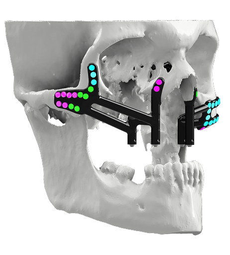
Imaging and Virtual Planning
The basis of virtual surgical planning is the high-resolution CT scan. Defective areas are outlined and 3D models with details of the defect are prepared to be used for preoperative planning. This yields valuable information for planning reconstruction of mandibular defects and maxillary defects treatment.
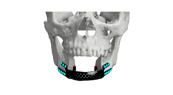
Reconstruction of Single-Sided Defects
For mandibular and maxillary resections, the unaffected side is digitally mirrored. This guarantees an ideal anatomical fit and provides for normal facial balance in mandibular reconstruction and maxillary reconstruction cases.
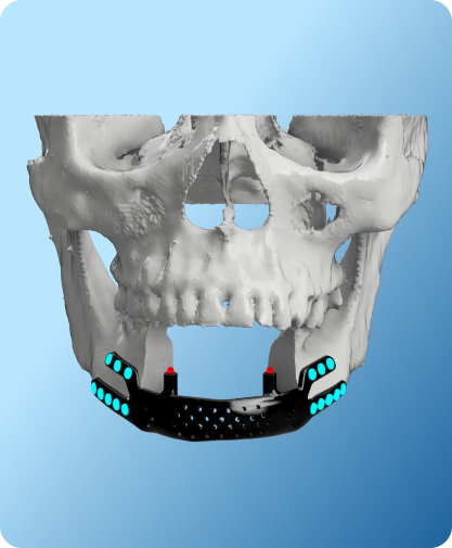
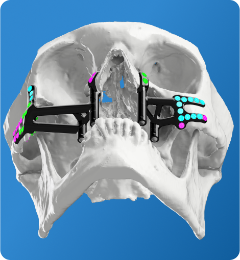
Base Plate and Fixation Design
A baseplate is then custom designed to fit the residual bone. Trajectory screws are well planed, without any passage to critical structures like canals or dental roots in order to ensure safety and stability.
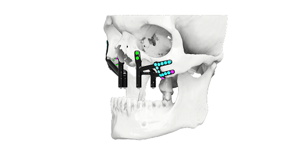
Biomechanical Optimization
Finite element analysis is performed before final fabrication for the evaluation of stresses and strains in the implant. Load bearing areas such as the mandibular angle, parasymphysis and condylar regions are provided with added strength. This mechanism guarantees biomechanical dependability for activities of mastication and speech, critical in trauma-related mandibular resection with the use of PSIs.
Clinical Applications of Patient-Specific Implants
Oncological Resection
For oral tumors and cancers, oncologic mandibular reconstruction with PSIs permits post-resection rehabilitation immediately. The prosthesis restores continuity of bone,supports the maxilla and orbital floor, reestablishing physiological as well as cosmetic function.

Infective Pathologies
There has been an increase in mucormycosis affected jaw reconstruction cases using 3D printing post-COVID-19. Patient-specific implants offer precise solutions for large defects, particularly in patients with high surgical risk of free flap surgery.
Trauma Reconstruction
Patient specific implants are used to fix complex maxillary and mandibular fractures and control bone loss. They re-establish occlusion, preserve facial balance and proportions without adding to intra-operative time.
Advantages of 3D-Printed Patient-Specific Implants
Anatomical Precision:
Precise fit through digital surgical planning and virtual mirroring.
Time Efficiency:
Prefabricated implants reduce surgical time compared to traditional methods.
Biomechanical Strength:
Finite element analysis in implants ensures long-term durability and
resistance to functional stresses.
Enhanced Functional and Aesthetic Outcomes:
Improved mastication, speech, and facial aesthetics.
Reduced Morbidity:
Avoids donor site complications of free flap surgery, making it ideal for
customized maxilla and mandible implants.
The application of 3D-printing using patient-specific implants in mandible and maxilla reconstruction represents a turn-around in craniofacial reconstruction. The integration of 3D printing, virtual surgical planning and biomechanical optimization of implants enable accurate anatomical restoration, functional rehabilitation, and excellent cosmetic results. 3D-printed patient specific implants restore the exact anatomy of maxilla and mandible and also improves the confidence and long-term oral health of patients.
FAQs
What is reconstruction of the resected mandible and maxilla?
It is a planned surgical process to restore jaw continuity, function, and facial form after tumour or cancer resection. Digital planning helps define resection margins and enables accurate reconstruction of the mandible and maxilla.
Why is pre-surgical planning important for mandible resection reconstruction?
Pre-surgical planning allows surgeons to visualize the defect in 3D, anticipate reconstruction challenges, and execute precise bone cuts. This improves accuracy, reduces operative time, and supports predictable functional outcomes.
How do mandible and maxilla reconstruction services support surgeons and hospitals?
These services integrate imaging, digital planning, and implant design into a single workflow, helping surgical teams perform complex reconstructions efficiently while maintaining hospital-grade standards and consistency.
What are the clinical advantages of PEEK implants in cranial reconstruction?What role do 3D printed jaw reconstruction implants play in surgery?
3D printed jaw reconstruction implants translate the digital plan into the operating room, allowing precise restoration of jaw anatomy and alignment, especially in complex oncologic defects.
What are patient-specific mandibular and maxillary implants?
Patient-specific mandibular and maxillary implants are custom-designed from the patient’s CT data to match individual anatomy, reducing intraoperative adjustments and improving fit and symmetry.
When are custom 3D printed mandibular and maxillary implants preferred?
Custom 3D printed mandibular implants and custom 3D printed maxillary implants are preferred in large, irregular, or complex defects where standard solutions may not restore anatomy accurately.
Can custom implants reduce recovery time after jaw reconstruction?
Yes ,custom implants tailored to individual anatomy can improve fit, reduce intraoperative adjustments, and support more accurate fixation. This often contributes to smoother rehabilitation and faster return to oral function than non-custom alternatives.
Contact Us & Get details for