- Work Hours : Mon to Sat : 09:30- 18:30


Doctor
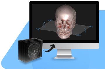
We use advanced software to preprocess images, such as noise reduction or contrast enhancement. This step ensures the input image is of the best quality for segmentation.
Doctor
We use advanced software to preprocess images, such as noise reduction or contrast enhancement. This step ensures the input image is of the best quality for segmentation.